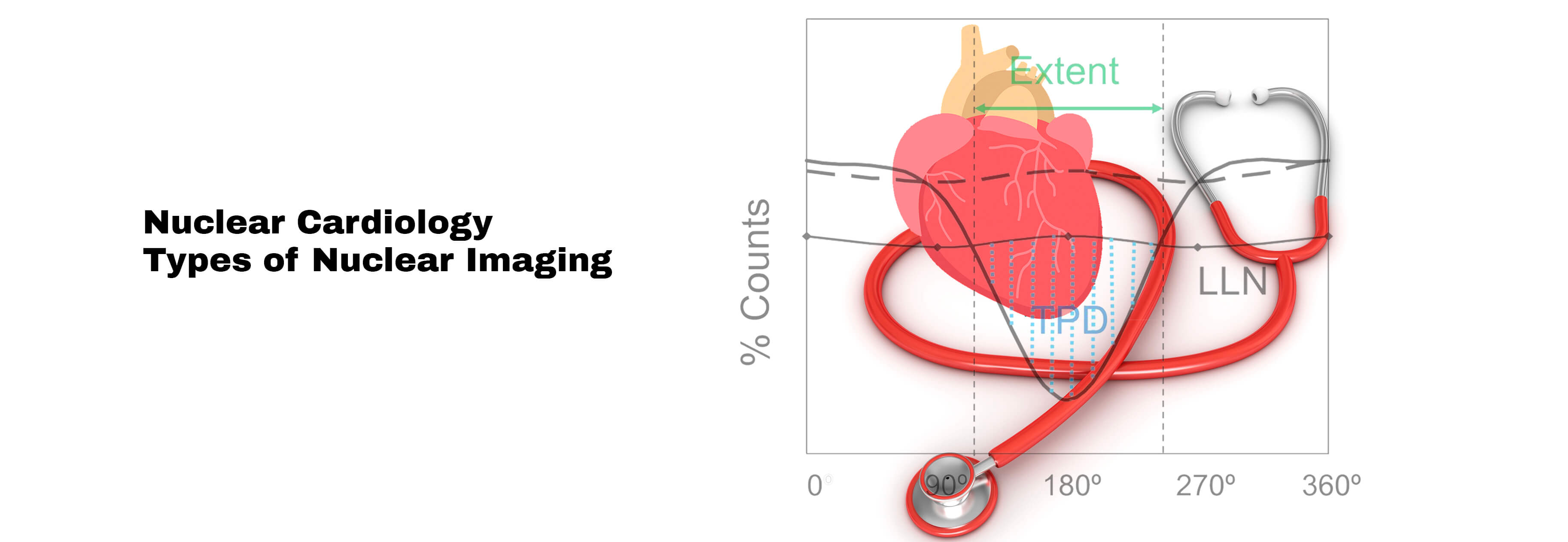
Nuclear Cardiology is nuclear cardiac imaging used to determine the adequacy of blood flow to the heart muscle during stress versus rest.
Its primary purpose is to identify the adverse effect of coronary artery disease or cardiomyopathy (a disease of the heart muscle) on the heart. Additionally, it can be used to determine whether the heart has been damaged by chemotherapy or radiation.
A radiotracer is a small amount of radioactive material injected into the bloodstream, inhaled, or swallowed by nuclear medicine practitioners.
To produce an image of the internal body organs, a special camera and computer detect gamma rays released by the radiotracer that travel through the area being examined.
The images produced by nuclear imaging medicine give unique information that cannot be obtained using other imaging forms.
Before sedation exam, an individual must inform the doctor about any recent illnesses, medical conditions, allergies, and medications currently used and if they are pregnant or breastfeeding.
So that doctor can advise about the appropriate variety and portion of the meal and before the exam, depending on the type of exam. Moreover, the patient should wear loose, comfortable clothes and put off jewelry. Wear patient gown if necessary.
Types of Nuclear Imaging:
- Equilibrium Radionuclide Angiogram (ERNA): This test gives insight to the doctor about the blood flow in the lower chambers.
- Exercise stress test with nuclear energy: This type of stress test helps doctors determine whether the heart is getting enough blood during exercise. During rest and an exercise session on a treadmill or stationary bike, technicians record the blood flow rate to the human heart.
- Pharmacological nuclear stress test: This test is a sim test, similar to a nuclear exercise stress test that administers medications to reduce the heart rate during exercise.
- An alternative to a pharmacological nuclear stress test, positron emission tomography (PET) uses different imaging devices and different radioactive materials. A few examples of these tests are:
- Cardiovascular nuclear imaging: Nuclear imaging helps the physician better understand the body’s organs and their functions. Additionally, it allows the doctor to visualize images of the heart, how it functions, and how well blood flows throughout the heart muscle.
Why do you need this test of Nuclear Cardiology?
Physicians use nuclear medicine studies for the diagnosis of cardiac disease includes;
- Unexplained chest pain
- Angina is triggered by exercise
- Exertion-induced breathlessness
- Electrocardiogram abnormalities
The following cardiac nuclear medicine procedures are performed;
- A myocardial perfusion scan visualizes the flow of blood to the heart walls.
- An evaluation is required to determine whether coronary artery disease is present or not.
- A heart attack or myocardial infarction causes severe damage to the heart.
- A procedure that restores blood supply to the heart after bypass surgery or another revascularization procedure.
- A cardiac gating technique is used to evaluate heart function and movement of the heart wall in conjunction with an electrocardiogram (ECG).
How to prepare for the test of Nuclear Cardiology?
An exam gown may be provided to the patient or allow for personal clothing. Before the test, the patient must inform the doctor about the following medical conditions or medication that can affect the test procedure and its result;
- Pregnancy or breastfeeding
- Allergies and illnesses experienced recently.
- Any medications you take, including vitamins and herbal supplement
- Myocardial infarction or heart attack
- Heart failure
- Asthma
- Bronchitis
- Lung adversity
- Abnormally conduction (like an AV block) of the heart, or aortic stenosis, or problems with the valves of your heart.
Additional General guidelines for Nuclear Cardiology
There are some other general instructions that patient should follow for the test;
- In addition, if you are experiencing problems with your knees, hips, or balance, you should let your doctor know as it could affect your ability to do the exercises.
- Make sure to wear comfortable clothes and shoes.
- The skin should not be oiled, moisturized, or creamed on the examination day.
- Don’t consume any caffeine (caffeinated and decaffeinated coffee, hot and cold tea, caffeinated soft drinks, energy drinks, chocolate, medications that contain caffeine, etc.) or smoke the 48 hours before your examination.
- Take medications with small amounts of water rather than eat or drink after midnight the day of your procedure.
- If you take nuclear medicine, ask your physician about their continuity or temporary discontinuation of beta-blockers or calcium channel blockers (Inderal, metoprolol, Norvasc, etc.).
During and after the test:
Nuclear medicine procedures are typically painless, except for intravenous injections. Significant side effects or discomfort are rare. During the needle insertion for the intravenous line, you will feel a slight pinprick.
The instructor may ask you to exercise until you are tired or short of breath or feel any discomfort that makes you stop, such as chest pain, leg pain, or shoulder pain.
If you cannot exercise, you may experience anxiety, dizziness, nausea, shakiness, or shaky sensations after taking a medication that increases blood flow.
Some people experience mild chest discomfort as well. After the infusion is complete, any symptoms that develop usually disappear. There are instances when other drugs can be given to make the side effects of medication less severe or less bothersome.
During the exam, you should remain calm as there is no pain associated with nuclear imaging. The only problem is that long periods standing or in one place may cause discomfort.
It is usually safe for you to resume your normal activities after your checkup unless your doctor instructs you for bed rest. Before you leave, you will receive special instructions from a nurse, technologist, or doctor for a healthy lifestyle and comfort.
Radioactive decay results in the loss of radioactivity over time in the small amount of radiotracer in your body. You should drink as much water as you can to flush out the material from your body.
What are the Risks of Nuclear Cardiology?
- When you take a drug for a stress test, you might experience chest pain if you have coronary artery disease. A physician will monitor your heart and, if needed, will administer medication.
- A cardiologist may consider same-day cardiovascular intervention if the test results suggest potentially life-threatening cardiac disease.
- Nuclear medicine exams expose you to a small amount of radiation using a small amount of radiotracer.
- Nuclear medicine has been used for diagnostics for almost six decades now. Low-dose exposure to such a chemical is not known to cause long-term ill effects.
- Before beginning, your doctor weighs the benefits and risks of any nuclear medicine treatment.
- It is infrequent for radiotracers to cause allergic reactions. If you are allergic to nuclear treatment, inform the nuclear medicine staff well before time.
References:
- https://www.radiologyinfo.org/en/info/cardinuclear retrieved on 8th December 2021.
- https://intermountainhealthcare.org/services/heart-care/treatment-and-detection-methods/nuclear-cardiology-tests/ retrieved on 8th December 2021.


